|
Previous Investigators
|
|
|
Kirsch Investigators 2003
|
|
Kirsch Investigators 2002
|
R.Clay Reid, MD, PhD
|
|
|
Kirsch Investigators 2001
|
|
|
|
|
|
Kirsch Investigators 2000
|
|
|
Ben A. Barres, MD, PhDKI 2000
Associate Professor of Neurobiology
Stanford School of Medicine
Biographical Sketch
Dr. Barres' major area of research is neuron-glial interactions in the developing central nervous system. He has pioneered the development of novel methods for the purification and culture of neurons and glial cells. Using these methods his lab has discovered that glial cells induce the formation of synapses as well as sodium channel clusters at nodes of Ranvier, both of which are necessary for rapid propagation of electrical signals in the brain. His lab has also discovered that electrical activity itself is necessary for the normal survival and growth of neurons. This has important implications for understanding how we learn as well as why the brain and spinal cord is not able to regenerate after injury.
Dr. Barres graduated from the Massachusetts Institute of Technology, obtained an MD degree at Dartmouth Medical School and completed his neurology residency at Cornell. He then obtained a PhD from Harvard Medical School and did his postdoctoral training at University College London, prior to joining Stanford in 1993.
Dr. Barres presently serves on the editorial boards of several leading journals including Neuron, the Journal of Neuroscience, and Development. He is a diplomate of the American Board of Psychiatry and Neurology. He has won many research awards including a Life Sciences Research Fellowship, the Klingenstein Fellowship Award, a McKnight Investigator Award, and a Searle Scholar Award, as well as teaching awards including the Kaiser Award for Excellence in Teaching and the Kaiser Award for Innovative and Outstanding Contributions to Medical Education. He presently serves on several advisory committees for the Society for Neuroscience, the National Multiple Sclerosis Society, and the National Institutes of Neurological Disorders.
Abstract of Research Plan
Provided by Dr. Barres in 2000
The functions of glial cells, which constitute about 90% of our brain's cells, are poorly understood. Currently, glia are thought to be primarily passive support cells that play little active role in the brain's cognitive abilities. We have recently discovered that glial cells secrete a protein that is essential for the formation of functional synapses between neurons in culture. We plan to identify this protein in order to investigate whether glial cells in vivo normally help to control synapse formation and function, thus actively participating in neural circuitry. We will use novel cell purification methods that we have developed to help separate the brain into its fundamental building blocks, defined types of neurons and glia. These methods provide a powerful tool, only newly available to neurobiologists, that has previously proved crucial to dissect cellular interactions in other complex tissues such as the immune system. We will then create transgenic mice that under- or over-express this protein in order to directly explore the role of glia in higher cognitive processes such as learning and memory. These studies are highly relevant to brain disorders such as stroke and neurodegenerative diseases such as Alzheimer's Disease, where glia become abnormal and may fail to perform their normal synaptic roles, thus contributing to cognitive impairment in these disorders. Investigation of the possible role of glia in cognitive function, learning, and memory is a new initiative by our lab made possible by funding from the Kirsch Foundation.
Publication Resulting from Foundation-Funded Work
E Ullian, S Sapperstein, K Christopherson, and BA Barres. 2001. Control of synapse number by glia. Science. 291: 657-61.
Back to top
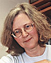
Elizabeth H. Blackburn, PhDKI 2000
Professor, Department of Biochemistry and Biophysics
Professor, Department of Microbiology and Immunology
University of California, San Francisco
Biographical Sketch
Dr. Elizabeth Blackburn is a leader in the area of telomere and telomerase research, and has broad experience in the different aspects of telomere function and biology. In 1985, she discovered the ribonucleoprotein enzyme, telomerase, and since that time, hers has become a lead laboratory in manipulating telomerase activity in cells. Having amassed considerable knowledge and experience in the effects this has on cells, Dr. Blackburn and her research team at the University of California, San Francisco are working with a variety of organisms and human cells, especially cancer cells, with the goal of understanding telomerase and telomere biology. Her work on telomeres and telomerase has been published extensively in peer-reviewed journals.
Dr. Blackburn earned her PhD in 1975 from the University of Cambridge in England, and did her postdoctoral work in Molecular and Cellular Biology from 1975 to 1977 at Yale. In 1978, Dr. Blackburn joined the faculty at the University of California, Berkeley in the Department of Molecular Biology. In 1990, she moved to University California, San Francisco (UCSF) and the Department of Microbiology and Immunology, where she served as Department Chair from 1993 to 1999. Dr. Blackburn is currently a Professor in the Department of Microbiology and Immunology, and the Department of Biochemistry and Biophysics at UCSF.
Dr. Blackburn is a foreign associate of the National Academy of Sciences (1993) and Past-President of the American Society for Cell Biology (1998). She is an elected fellow to the American Academy of Arts and Sciences (1991), the Royal Society of London (1992), the American Academy of Microbiology (1993), and the Institute of Medicine (2000). She received the Benjamin Franklin medal in Life Science (2005).
Abstract of Research Plan
Provided by Dr. Blackburn in 2000
Telomeres are the specialized natural ends of eukaryotic chromosomes. Functional ("capped") telomeres allow cells to proliferate, and protect chromosomes from genomic instability. The planned research arises directly from recent advances that followed up from our discovery and subsequent analysis of telomerase. The ribonucleoprotein telomerase synthesizes telomeric DNA and is required for long-term telomere maintenance. Telomerase is commonly activated in human cancer cells, but not in most normal somatic cells. Manipulating telomerase activity has potential therapeutic uses, both in activating the proliferation of normal cells in order to renew aging human tissues, and in treating cancer.
The proposed research explores fundamental and applied aspects of telomerase action. The first part tests specific hypotheses emerging from our new conceptualization of telomere function. Does telomerase act in response to DNA damage signals from uncapped telomeres, and itself signal cells to proliferate? How does telomerase contribute to telomere capping? The second, closely linked part of the research exploits the activation of telomerase in cancer cells. We are developing a promising novel anti-cancer therapy, a gene "anti-therapy" approach based on interfering with telomere function. We found that expressing various mutant forms of telomerase engineered to synthesize deleterious telomeric sequences ("toxic" telomeres), greatly slows growth of human cancer cells, and induces their suicide. We showed such a strategy works effectively and rapidly in lower eukaryotes because the "toxic" telomeres block chromosome separation and hence cell division. I will investigate the mechanisms underlying growth effects in human cancer cells.
Publications Resulting from Foundation-Funded Work
EH Blackburn. 2001. Switching and signaling at the telomere. Cell. 106: 661-673.
S Chan, J Chang, J Prescott, and EH Blackburn. 2001. Altering telomere structure allows telomerase to act in yeast lacking ATM kinases. Current Biology. 11: 1240-1250.
S Chan and EH Blackburn. 2001. New ways not to make end meet: telomerase, DNA damage proteins and heterochromatin. Oncogene. 21: 553-563.
MM Kim, MA Rivera, IL Botchkina, R Shalaby, AD Thor, and EH Blackburn. 2001. A low threshold level of expression of mutant-template telomerase RNA is sufficient to inhibit human tumor cell growth. Proc. Natl. Acad. Sci. U.S.A. 98: 7982-7987.
SWL Chan and EH Blackburn. 2003. Telomerase and ATM/Tel1p Protect Telomeres from Nonhomologous End Joining. Molecular Cell. 11: 1379-1387.
B Williams and AJ Lustig. 2003. The Paradoxical Relationship between NHEJ and Telomeric Fusion. Molecular Cell. 11: 1125-1126.
Back to top
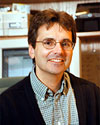
Richard T. Born, MDKI 2003
Associate Professor of Neurobiology
Harvard Medical School
Biographical Sketch
A large portion of the primate brain is dedicated to "seeing." Dr. Born's lab uses this important sense as a model system for studying how the brain works. In particular, Dr. Born and his colleagues are trying to decipher the neural circuitry responsible for our ability to perceive visual motion and to track moving targets with our eyes. In what direction is an object moving? How fast? Is it really moving with respect to its surroundings, or does it just seem to be moving because I'm moving? The calculations that allow our brains to answer these critical questions are performed by populations of neurons that are anatomically arranged to form a map representing all possible directions of motion in all parts of the visual field. Certain comparative processes, such as those that allow us to distinguish a moving object from the visual background, appear to be performed by a subset of motion-sensitive neurons whose receptive fields have opponent surrounds. Ongoing projects in the lab are designed to understand precisely how this receptive field property is related to motion perception.
Because many people afflicted with schizophrenia have specific deficits in both motion perception and visually-guided eye movements, the researchers in Dr. Born's lab are also attempting to understand how the circuitry they study in animals may be related to this human disorder. To this end, they study the effects of dopamine and dopaminergic drugs on cortical motion processing circuits and motion perception. It is anticipated that manipulating this neuromodulator will allow them to reproduce some of the clinical findings in an animal model, where they can unravel the underlying mechanisms. Conversely, some of Dr. Born's discoveries from animal studies are being used to design visual tests for people with schizophrenia.
Dr. Born received his B.A. summa cum laude in Chemistry from DePauw University in 1983 and his M.D. from Harvard in 1988. He continued postdoctoral training at Harvard Medical School with Drs. David Hubel and Margaret Livingstone from 1988 to 1991 and at Stanford University Medical School with Dr. William Newsome from 1992 to 1994. He moved back to Boston in 1995 as an Assistant Professor of Neurobiology at Harvard Medical School and was promoted to Associate Professor in 2001.
Dr. Born's past honors include a Klingenstein Fellowship in the Neurosciences, a Physician Scientist Award from the National Eye Institute, and a Whitehall Foundation Award.
Abstract of Research Plan
Provided by Dr. Born in 2003
Many schizophrenics and their first-degree relatives have specific deficits in smooth pursuit eye movements and in motion perception. Both of these deficits are similar to those seen with lesions in a specific cortical region, known as the middle temporal visual area (MT), of macaque monkeys. A finer, circuit-level, link involves the chemical dopamine. This neuromodulator is strongly implicated in the pathophysiology of schizophrenia, and it causes an interesting alteration in a specific kind of brain circuit, referred to as "center-surround opponency," that abounds in MT and that has been a major focus of the work in my laboratory. Because the center-surround interactions in MT can be directly related to specific roles in both motion perception and eye movements, we propose to study the effects of dopamine on these neurons in alert monkeys while simultaneously recording the monkeys' eye movements. This will enable us to not only characterize dopaminergic effects on the local circuits that account for the receptive fields of cortical neurons, but to directly relate these effects to alterations in behavior. An additional important element in our approach is the use of computational models of MT circuitry. These models make specific and testable predictions concerning the effects on eye movements caused by alterations in the balance between center and surround of the receptive fields of single neurons. Thus the long-term promise of the proposed experiments is to build a bridge from neurophysiology to schizophrenia using pharmacology, cortical motion processing, smooth pursuit eye movements and computational modeling as tools.
Publications Resulting from Foundation-Funded Work
CC Pack, JN Hunter, and RT Born. 2005. Contrast dependence of suppressive influences in cortical area MT of alert macaque. J Neurophysiology. 93: 1809-1815
RT Born, CC Pack, and JN Hunter. 2005. Modulation of center-surround interactions in cortical visual area MT of the alert macaque monkey. Abstract presented at the 2005 International Congress on Schizophrenia Research, April 2-6, 2005, Savannah, GA.
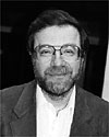
Ronald A. DePinho, MDKI 2000
American Cancer Society Research Professor
Professor of Medicine and Genetics
Harvard Medical School;
Director of Transgenesis and Gene Targeting
Dana Farber Cancer Institute
Biographical Sketch
Dr. DePinho has been a leader in the use of engineered mouse models to dissect the molecular mechanisms and biological processes governing the genesis of cancer. These studies have provided insight into how common molecular changes in aspiring cancer cells influence cellular growth and survival, allowing for acquisition of the fully transformed state. Dr. DePinho and his colleagues have also produced cancer models that permit an analysis of the complex symbiotic host-tumor cell interactions required for tumor maintenance. These changes also impinge on pathways integral to the development of chronic diseases afflicting many millions worldwide – diseases such as liver cirrhosis and liver cancer. Dr. DePinho has organized efforts to disseminate transgenic and gene targeting technologies to many investigators worldwide.
Dr. DePinho studied Art History and Biology at Fordham University where he graduated with honors. He received his MD degree with distinction from the Albert Einstein College of Medicine. His independent research career began at the Albert Einstein College of Medicine where he was the Senior Scholar in Cancer Research. He is currently a member of the Department of Medicine and Genetics of Harvard Medical School and the Department of Adult Oncology at the Dana Farber Cancer Institute. Dr. DePinho's honors and awards include the James S. McDonnell Scholar Award, the Cancer Research Institute Scholar Award, the Melini Award for Biomedical Excellence, the Irma T. Hirshl Award, and the 2002 American Society of Clinical Investigation Award. He is an American Cancer Society Research Professor.
Abstract of Research Plan
Provided by Dr. DePinho in 2000
Liver cirrhosis and hepatocellular carcinoma are among the leading causes of death in humans. Diverse etiologies, ranging from chronic viral infections to alcohol abuse, all act to invoke continual cycles of hepatocyte destruction and regeneration. Ultimately, terminal liver failure ensues as hepatocyte lose proliferative potential and extensive fibrosis replaces the liver parenchyma. Mechanisms underlying these signal events are not understood. Recently, we provided evidence that telomere attrition, brought about by chronic cycles of hepatocyte death and regeneration, precipitates a pathogenetic chain of events that leads to end-stage liver failure, proliferative arrest, and fibrosis. Moreover, short telomeres in human cirrhotic livers is consistent with our hypothesis. Strikingly, somatic telomerase reconstitution in the livers of telomerase-deficient mice blocked experimental cirrhosis and improved survival. This entirely new line of research provides the first genetic model for the study of liver cirrhosis and somatic telomerase therapy. Here our efforts will be directed towards the characterization of new telomerase reverse transcriptase (TERT) knockout mouse that is ideal for the evaluation of telomerase therapy. In these studies, TERT null mice will be subjected to genetic, surgical and toxin-mediated protocols designed to test the regenerative potential and cirrhotic propensity of the livers of mice with telomere dysfunction. Next, several gene- and protein-based delivery methods will be tested for restoration of telomere function and block the cirrhotic response. These studies will allow us to ascertain the anti- and pro-cancer properties of somatic telomerase reconstitution and the role of telomere dysfunction in hepatocellular carcinogenesis.
Publications Resulting from Foundation-Funded Work
KL Rudolph, S Chang, M Millard, N Schreiber-Agus, and RA DePinho. 2000. Inhibition of experimental liver cirrhosis in mice by telomerase gene delivery. Science. 287:1253-1258.
SE Artandi, S Chang, SL Lee, S Alson, GJ Gottlieb, L Chin, and RA DePinho. 2000. Telomere dysfunction promotes non-reciprocal translocations and epithelial cancer in mice. Nature. 406:641-645.
KK Wong, S Chang, SR Weiler, S Ganesan, J Chaudhuri, C Zhu, SE Artandi, KL Rudolph, GJ Gottlieb, L Chin, FW Alt, and RA Depinho. 2000. Telomere dysfunction impairs DNA repair and enhances sensitivity to ionizing radiation. Nat Genet. 26:85-8.
SE Artandi and RA DePinho. 2000. A critical role for telomeres in suppressing and facilitating carcinogenesis. Curr Opin Genet Dev. 10: 39-46.
SE Artandi and RA DePinho. 2000. Mice without telomerase: what can they teach us about human cancer? Nature Medicine. 6:4-7.
KH Lee, KL Rudolph, YJ Ju, RA Greenberg, L Cannizzaro, L Chin, SR Weiler, and RA DePinho. 2001. Telomere dysfunction alters the chemotherapeutic profile of transformed cells. Proc Natl Acad Sci USA. 98:3381-3386.
KL Rudolph, M Millard, MW Bosenberg, and RA DePinho. 2001. Telomere dysfunction and evolution of intestinal carcinoma in mice and humans. Nature Genetics. 28:155-158.
MT Hemann, KL Rudolph, MA Strong, RA DePinho, L Chin, and CW Greider. 2001. Telomere dysfunction triggers developmentally regulated germ cell apoptosis. Molecular Biology of the Cell. 12:2023-30.
V Ranganathan, WF Heine, DN Ciccone, KL Rudolph, X Wu, S Chang, H Hai, IM Ahearn, DM Livingston, I Resnick, F Rosen, E Seemanova, P Jarolim, RA DePinho, and DT Weaver. 2001. Rescue of a telomere length defect of Nijmegen breakage syndrome cells requires NBS and telomerase catalytic subunit. Curr Biol. 11:962-6.
S Chang, C Khoo, and RA DePinho. 2001. Modeling chromosomal instability and epithelial carcinogenesis in the telomerase-deficient mouse. Seminars in Cancer Biology. 11:227-39.
Kucherlapati and RA DePinho. 2001. Cancer: Telomerase meets its mismatch. Nature. 411:647-8.
S Chang and RA DePinho. 2002. Telomerase Extracurricular Activities. Proc Natl Acad Sci. 99:12520-2.
RS Maser and RA DePinho. 2002. Keeping telomerase in its place. Nat Med. 8:934-6.
SE Artandi, S Alson, MK Tietze, NE Sharpless, S Ye, RA Greenberg, DH Castrillon, JW Horner, SR Weiler, RD Carrasco, and RA DePinho. 2002. Constitutive telomerase expression promotes mammary carcinomas in aging mice. Proc Natl Acad Sci. 99:8191-8196.
RC O'Hagan, S Chang, RS Maser, R Mohan, SE Artandi, L Chin, and RA DePinho. 2002. Telomere dysfunction provokes regional amplification and deletion in cancer genomes. Cancer. 2:149-155.
S Chang, CM Khoo, ML Naylor, RS Maser, and RA DePinho. 2003. Telomere-based crisis: functional differences between telomerase activation and ALT in tumor progression. Genes and Dev. 17(1):88-100.
KK Wong, RS Maser, RM Bachoo, J Menon, DR Carrasco, Y Gu, FW Alt, and RA DePinho. 2003. Telomere dysfunction and ATM deficiency deplete stem cell reserves and accelerate aging. Nature. 4421:(6923):643-8.
Book Chapter:
KL Rudolph and RA DePinho. Telomeres and telomerase in experimental liver cirrhosis. In: The Liver: Biology and Pathobiology. Fourth Edition. IM Arias, JL Boyer, EV Chisari, N Fausto, D Schachter, and D Shafritz (ed.), Lippincott Williams & Wilkins: Philadelphia 2001; pp. 999-1009.
Allison J. Doupe, MD, PhDKI 2001
Professor of Psychiatry & Physiology
And the Keck Center for Integrative Neuroscience
University of California, San Francisco
Biographical Sketch
Dr. Doupe's area of research is the learning of complex vocal behavior in the songbird, a powerful model system for the study of how the nervous system generates behavior, both normally and after altered experience or brain injury. Her lab has shown that the properties of neurons in a basal ganglia circuit of the songbird brain are shaped both by the sound of the bird's own voice as it learns and by the sound of the tutor that it is copying, and that this circuit is active during singing and required for song plasticity throughout the birds' life. Additional experiments have revealed that brain activity in these areas is influenced by the social context, and have begun to explore the cellular mechanisms of learning. These studies have important implications for understanding how the brain learns, with particular relevance to the learning of human speech.
Dr. Doupe received her B.Sc. at McGill University, Canada, and then graduated with a Ph.D. and an M.D. from the Harvard-MIT M.D. Program in Health Sciences and Technology and the Harvard Medical School Neurobiology Department. Her graduate work, with Paul Patterson, Ph.D., investigated the effects of growth factors and steroid hormones on neural development. After a medicine internship at Massachusetts General Hospital in Boston, and psychiatry residency at the University of California, Los Angeles, Neuropsychiatric Institute, she carried out postdoctoral work on the neurophysiology of the songbird brain at the California Institute of Technology, with Mark Konishi. In 1993 she moved to the Departments of Psychiatry and Physiology and the Keck Center for Integrative Neuroscience at UCSF, where she is currently an Associate Professor.
She is the recipient of many research awards including a Klingenstein Fellowship, a McKnight Investigator Award, a Searle Scholarship, an EJLB Scholar Award, and a Merck Fellowship. She is a diplomate of the American Board of Psychiatry and Neurology, and serves on the editorial board of several leading journals, including the Journal of Neuroscience and the Journal of Neurobiology.
Abstract of Research Plan
Provided by Dr. Doupe in 2001
Cortical-basal ganglia (C-BG) circuitry is critical for motor and reinforcement learning and an important site of dysfunction in neurological and psychiatric disorders including Parkinson's disease. Greater basic knowledge of the functions of this circuitry and of its dopamine (DA) innervation will be crucial for making progress in understanding learning as well as C-BG diseases. In this regard, songbirds provide a unique animal model: they have specialized a portion of their C-BG circuitry for learning and production of their stereotyped vocal motor behavior. This simplifies the task of making direct causal links between C-BG circuit activity and behavior, and gives this system the potential to provide insight into the general computations carried out by C-BG pathways and their DA inputs in all vertebrates. To this end, we propose two research directions, which extend our previous work. 1) We will simultaneously record neural activity from the song C-BG circuit and its target motor nucleus in awake, behaving animals at different stages of learning, both in normal birds and in birds where we have experimentally disrupted the activity of this circuit. This will directly test how the patterns of activity in the C-BG pathway relate to behavior and influence motor circuitry. 2) We will investigate the role of DA in this C-BG circuit by directly studying the release of DA and its effect on the circuitry, and by blocking or depleting DA inputs to this pathway (thus mimicking disease) in adults and during learning, and assessing the effects on neural activity and behavior.
Publications Resulting from Foundation-Funded Work
EC Ihle, GD Carrillo, M Giorgetti, L Tecott, and AJ Doupe. 2003. Measurement and manipulation of dopamine in the awake, behaving zebra finch basal ganglia. Soc Nsci Abstr.
MH Kao, AJ Doupe, and MS Brainard. 2003. Lesions of LMAN prevent context-dependent changes to song variability. Soc Nsci Abstr.
EC Ihle, GD Carrillo, M Hu, JB Becker, and AJ Doupe. 2004. Modulation of male zebra finch striatal dopamine levels by courtship behavior. Soc Nsci Abstr.
MH Kao and AJ Doupe. 2004. Context-dependent differences in the activity of single units in a basal ganglia-forebrain circuit required for song plasticity. Soc Nsci Abstr.
Peter K. Jackson, PhDKI 2003
Associate Professor of Pathology and Microbiology and Immunology
Stanford University School of Medicine
Biographical Sketch
Dr. Jackson's work focuses on critical enzymatic mechanisms controlling eukaryotic cell growth. He has made also made important contributions to understanding how non-catalytic domains are used to target enzymes to their intracellular sites of action. His graduate work with David Baltimore at MIT contributed to our understanding of how tyrosine kinases use modular domains, called SH2 and SH3, to target tyrosine phosphorylation of specific targets and how nuclear localization is regulated. During his postdoctoral studies with Marc Kirschner at UCSF and Harvard Medical School, and continuing in his own laboratory at Stanford, Dr. Jackson defined mechanisms by which cyclin-dependent kinases are activated selectively at origins of DNA replication and how ubiquitin ligases are selectively activated. At Stanford, Dr. Jackson has studied the role of ubiquitin ligases, cyclin-dependent kinases, and regulatory phosphatases to control the chromosome and centrosome cycles. This work led to the discovery of the role of cyclin-dependent kinases and ubiquitin ligases in the centrosome duplication cycle (with Tim Stearns of Biological Sciences), the role of the Cdc14 phosphatase in controlling cytokinesis in animal cells, and the discovery of a new class of regulators of ubiquitin ligases and the biochemical mechanism by which they work. Work on the Emi1 regulator of ubiquitin ligases has led to fundamental insights into the control of mitosis and provided a critical link between a genetically defined pathway controlling cell growth and regulators of cell division.
Dr. Jackson received a BA in Mathematics and in Economics Magna cum Laude from Yale College. He worked at the RCA Corporation as a mathematician and computer scientist before pursuing graduate work in Chemical Physics at the University of Chicago and receiving a PhD in Biophysics from Harvard University. After postdoctoral work, he joined the faculty at Stanford. At Stanford, Dr. Jackson teaches courses in chemical biology, cell biology, biochemistry, and on the molecular basis of cancer. Dr. Jackson is a consultant to the pharmaceutical and biotechnology industries, a member of the editorial board of Developmental Cell, and has served on NIH study sections. He was a Merck Fellow of the Life Sciences Research Foundation, a Baxter Scholar, a Lutje-Stubbs Scholar, and the William T. Cohen Memorial Lecturer at the Dana-Farber Cancer Institute.
Abstract of Research Plan
Provided by Dr. Jackson in 2003
Regulated destruction of substrates of SCF ubiquitin ligases is critical for the control of processes ranging from the cell cycle and development to signaling and homeostatic control in adults. The ability of the adapter subunits of SCF ubiquitin ligases to recognize phosphorylated peptide sequences in their target proteins is typically the triggering step for destruction of those targets. These adapter proteins are called "F-box" proteins and the two major classes use either a WD40 repeat/ b-propeller domain or a leucine rich repeat domain as the scaffold for substrate recognition. A new crystal structure of an F-box WD40 proteins (Fbw) bound to its phosphorylated substrate provides a rationale for the specificity of how these phosphophorylated target sequences, called phosphodegrons, is achieved. The structure further provides a model to highlight the specificity determining sequences on specific adapter proteins and to help decode new phosphodegrons in the mammalian genome. This grant is directed toward using this new information to systematically determine the phosphodegron code and to create new affinity probes for measuring the activity of these destruction pathways in situ. These in situ probes will be useful for both research and diagnostics applications and have the potential to be vastly more informative than probing message or protein levels alone. We plan to apply these technologies to applications in cancer and neurodegeneration.
Publications Resulting from Foundation-Funded Work
PK Jackson and AG Eldrige. 2002. The SCF Ubiquitin Ligase: An Extended Look. Molecular Cell 9, May.
AGM Borchers, AL Hufton, AG Eldrige, PK Jackon, RM Harland, and JC Baker. 2002. The E3 ubiquitin ligase GREUL1 causes ectopic cement gland development in Xenopus embryos. Developmental Biology. 251: 395-408.
D Hansen, J Hsu, A Eldridge, B Kaiser, and PK Jackson. 2002. Control of the centriole and centrosome cycles by ubiquitin ligases. Oncogene. 21: 6209-6221.
L He, XY Lu, AF Jolly, AG Eldridge, SJ Watson, PK Jackson, GS Barsh, and TM Gunn. 2003. Spongiform degeneration in mahoganoid mutant mice. Science. 299: 710-723.
PK Jackson. 2003. Ubiquitinating a Phosphorylated Cdk Inhibitor on the Blades of the Cdc4 beta-Propeller. Cell. 112: 142-4.
F Margottin-Goguet, JY Hsu, A Loktev, HM Hsieh, JDR Reimann, and PK Jackson. 2003. Destruction of Emi1 in late prophase requires the SCFrCP/Slimb ubiquitin ligase and triggers activation of the Anaphase Promoting Complex to allow progression beyond prometaphase. Developmental Cell. 4: 813-26.
D Guardavaccaro, Y Kudo, J Boulaire, M Barchi, L Busino, M Donzelli, F Margottin-Goguet, PK Jackson, L Yamasaki, and M Pagano. 2003. Control of Meiotic and Mitotic Progression by the F Box Protein beta-Trcp1 in Vivo. Developmental Cell. 4: 799-812.
PK Jackson. 2004. An Introduction to Posttranslational Control by Ubiquitiin-dependent Proteolysis. In Inborn Error of Development, Oxford Monographs on Medical Genetics. No. 49, Oxford.
PK Jackson. 2004. Linking tumor suppression, DNA damage and the anaphase-promoting complex. Trends Cell Biol. 14: 331-4.
DV Hansen, A Loktev, K Ban, and PK Jackson. 2004. P1k1 Regulates Activation of the Anaphase Promoting Complex by Phosphorylating and Triggering SCF?TrCP –Dependent Destruction of the APC Inhibitor Emi1. Mol Biol Cell. 15: 5623-5634.
Alex L. Kolodkin, PhDKI 2002
Associate Professor of Neuroscience
The Johns Hopkins University School of Medicine
Biographical Sketch
Dr. Kolodkin's research focuses on how neurons find their targets during development. Working in both invertebrates (Drosophila and grasshopper) and vertebrates (mouse and rat), his laboratory has examined the cellular and molecular mechanisms which underlie repulsive axon guidance. Dr. Kolodkin and his colleagues have defined the repulsive effects of proteins belonging to the semaphorin family, identified semaphorin receptors, and most recently have found novel signaling components critical for semaphorin-mediated repulsion. Many of these observations have important clinical implications for the promotion of neuronal regeneration following injury.
Dr. Kolodkin received his B.A. in Biological Sciences and Religious Studies from Wesleyan University in Middletown, Connecticut. He obtained graduate training at the Institute of Molecular Biology at the University of Oregon, where he received his Ph.D. in Molecular Biology and Genetics for work on molecular mechanisms of genetic recombination. Dr. Kolodkin did post-doctoral work in neural development in the Department of Molecular and Cell Biology at the University of California, Berkeley, prior to joining the Department of Neuroscience at The Johns Hopkins School of Medicine in 1995. Dr. Kolodkin's honors include Scholar and Investigator Awards from the Howard Hughes Medical Institute, the McKnight Foundation, a Klingenstein Fellowship Award, a Searle Scholars Award, a Sloan Research Fellow Award, and a Whitehall Research Award. Dr. Kolodkin serves on the Editorial Advisory Board of the journal Neuron and is also an Advisory Board Member of the New York State Spinal Cord Injury Research Foundation
Abstract of Research Plan
Provided by Dr. Kolodkin in 2002
The goal of this project is to provide insight into neuronal guidance mechanisms in order to discover methods of attenuating repulsive guidance–an essential step on the road toward promoting therapeutic neuronal regeneration. Repulsive guidance cues, like attractive cues, play key roles in directing axons to their targets during neural development. Many repulsive guidance mechanisms are likely to function in the adult nervous system as well–preventing neuronal regeneration following injury or neurodegenerative events. We propose here exploiting the advantages of revealing these mechanisms in a model invertebrate system, the fruit fly Drosophila, and then directly applying this understanding to experiments directed towards analysis in mammalian neurons. Specifically, with support from the Kirsch Foundation, we will conduct genetic screens in Drosophila in order to directly identify novel signaling pathways used by neurons in response to members of the semaphorin family of growth cone guidance cues–a large phylogenetically conserved family of axonal repellents. We will then directly address how these same signaling components direct semaphorin-mediated repulsion in mammalian neurons. A specific aim of this project is an in-depth analysis of a novel, phylogenetically conserved, family of proteins we have recently shown is required for semaphorin receptors to mediate axonal repulsion. These proteins contain domains which suggest a novel mechanism by which neurons may be repelled. We will use genetic approaches in mice, neuronal leasion paridigms in rats, and pharmacological approaches in neuronal cell culture to establish how inhibition of these semaphorin signaling components might promote neuronal regeneration.
Publications Resulting from Foundation-Funded Work
C Gu, DV Reimert, T Shu, B Fritzsch, LJ Richards, ER Rodriquez, AL Kolodkin, and DD Ginty. 2003. Neurophilin-1 integrates semaphoring and VEGF signaling during neural and cardiovascular development. Developmental Cell. 5: 45-57.
A Sahay, ME Molliver, DD Ginty, and AL Kolodkin. 2003. Semaphorin 3F is critical for limbic system connectivity and is required in neurons for selective CNS axon guidance events. J. Neuroscience. 23: 6671-6680.
RJ Pasterkamp, JJ Peshon, MK Spriggs, and AL Kolodkin. 2003. Semaphorin 7A promotes axon outgrowth through integrins and MAPKs. Nature. 424: 398-405.
JR Terman and AL Kolodkin. 2004. The AKAP Nervy direction couples Protein Kinase A to Plexin-mediated semaphoring repulsion. Science. 303: 1204-1207.
AB Huber, AL Kolodkin, DD Ginty, and JF Cloutier. 2003. Signaling at the growth cone: Ligand-receptor complexes and the control of axon growth and guidance. Ann Rev Neuroscience. 26: 509-563.
RH Pasterkamp and AL Kolodkin. 2003. Semaphorin Junction: making tracks toward neuronal connectivity. Curr Opin Neurobiology. 13: 79-89.
Wendell A. Lim, PhDKI 2002
Associate Professor of Cellular and Molecular Pharmacology, and of Biochemistry and Biophysics
University of California, San Francisco
Biographical Sketch
Dr. Lim's research focuses on how eukaryotic signal transduction pathways are wired. His lab is utilizing structural, biochemical, and cell biological methods to understand how proteins involved in signal transduction are able to process biological information in manner that maintains high specificity, but also the ability to integrate complex inputs. His lab is studying several key proteins in pathways that regulate cell growth, stress responses, and movement.
Dr. Lim obtained his Ph.D. in 1991 from the Massachusetts Institute of Technology. He did his postdoctoral work at Yale University before joining the faculty at the University of California, San Francisco in 1996.
Dr. Lim's honors include a Burroughs Wellcome Young Investigator in the Pharmacological Sciences Award, a Searle Scholar Award, and a Fellowship for Science and Engineering from The David and Lucile Packard Foundation.
Abstract of Research Plan
Provided by Dr. Lim in 2003
Living cells contain complex networks of signaling proteins that allow them to respond appropriately to environmental cues. A major question in biology is how cell signaling occurs with high specificity. Given the vast number of stimuli, signaling proteins, and possible responses, how is proper information flow normally maintained? Inversely, in disease states such as cancer, why are improper signals transmitted?
An emerging paradigm is that scaffold proteins play an important role in maintaining signaling specificity. Scaffold proteins interact with multiple proteins in a pathway, and are hypothesized to constrain these proteins to efficiently interact only with one another and not with related proteins excluded from the scaffolded complex. Scaffold proteins are found in many different cell types, ranging from yeast to mammalian neurons, and they play a role in diverse types of protein pathways. Despite the phenomenological importance of scaffold proteins, remarkably little is known about the biochemical mechanisms by which they control cellular information flow. Our overall goal is to understand the molecular basis by which scaffolds control signaling efficiency and specificity by undertaking a biochemical and structural analysis of model scaffolding proteins involved in mitogen activated protein (MAP) kinase pathways in yeast. These studies will shed light on how information flow is controlled in cells, and may reveal new avenues by which we can manipulate these circuits in the case of signaling-based diseases.
Publication Resulting from Foundation-Funded Work
A Zarrinpar, RP Bhattacharyya, MP Nittler, and WA Lim. 2004. Sho1 and Pbs2 Act as Coscaffolds Linking Components in the Yeast High Osmolarity MAP Kinas Pathway. Molecular Cell. 14: 825-832.
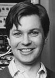
R. Clay Reid, MD, PhDKI 2002
Associate Professor of Neurobiology
Harvard Medical School
Biographical Sketch
Dr. Reid has made numerous contributions to the understanding of visual processing in the mammalian brain. As signals are transmitted along the pathway from the retina, through the thalamus, and on to the cerebral cortex, there is a profound transformation of how visual information is encoded. Neurons at all three levels respond to a very limited range of stimuli: they fire action potentials when presented with a stimulus whose luminance or color changes in a specific way over time or across the visual scene. In particular, visual cortical neurons are highly selective for the orientation of a stimulus and its direction of movement, although the thalamic neurons that provide input to the cortex are non-selective. Dr. Reid has employed a number of advanced experimental and analytical techniques to help unravel how this profound transformation takes place. By making electrical recording from multiple neurons simultaneously in retina, thalamus and/or visual cortex, she has helped trace the exquisitely precise circuitry responsible for the critical first stages in visual processing. Dr. Reid has contributed to our knowledge of many aspects of this pathway, including the processing of motion, orientation, and color.
Dr. Reid Received a B.S. summa cum laude in Mathematics and in Physics and Philosophy from Yale University in 1982. He received a Ph.D. from the Rockefeller University in 1988 and an M.D. from Cornell University Medical College in 1991. He did postdoctoral work with Robert Shapley at New York University and in Torsten Wiesel's laboratory at the Rockefeller University, where he was promoted to Assistant Professor in 1993. In 1996, he moved to the Department of Neurobiology at Harvard Medical School, where he is currently Associate Professor.
Dr. Reid's past honors include a Klingenstein Fellowship in the Neurosciences and the Young Investigator Award from the Society for Neuroscience.
Abstract of Research Plan
Provided by Dr. Reid in 2003
For those who have lost vision due to accident or to degenerative retinal diseases, restoration of visual function would mean a tremendous increase in the quality of life. Spurred by the success of cochlear implants—which have provided limited hearing to patients with several forms of deafness—there has been a resurgence of work on devices to stimulate the visual system electrically. Most recent work on visual prostheses has been aimed at developing intra-ocular devices for retinal stimulation. We plan instead to stimulate the lateral geniculate nucleus (LGN), the part of the thalamus that relays signals from the retina to primary visual cortex.
Our approach will be to implant bundles of microscopic electrodes in the LGN of animals that can later be studied in the alert state. These electrodes can be used both to record the normal activity of LGN neurons and to stimulate the neurons electrically. This project combines two lines of research from our laboratory: multi-electrode recordings from neural ensembles in the LGN, and studies of visual cortical responses to electrical stimulation of the LGN. The advantage of working in alert animals is that we can examine the physiological responses of visual cortical neurons at the same time that we explore the perceptual correlates of electrical stimulation. The long-term goal is to restore low-resolution but usable visual percepts in otherwise blind or low-vision patients. Given the current state of brain-stimulation technologies and our strong understanding of the early visual system, this goal should now be within reach.
Publication Resulting from Foundation-Funded Work
P Kara, JS Pezaris, S Yurgenson, and RC Reid. 2002. The spatial receptive field of thalamic inputs to single cortical simple cells revealed by the interaction of visual and electrical stimulation. Proc Natl Acad Sci USA. 99: 16261-6.
Geraldine C. Seydoux, PhDKI 2001
Associate Professor of Molecular Biology & Genetics
Johns Hopkins University School of Medicine
Biographical Sketch
Dr. Seydoux's research focuses on the development of the germline (cells that become the eggs and sperm in humans). She is investigating the mechanisms that lead to the establishment of germline stem cells during early development and has found that global inhibition of mRNA transcription is an essential first step in the establishment of the germline. Her lab has also shown that members of an ancient gene family conserved throughout evolution (nanos-related genes) are essential to maintain the viability and totipotency of germline stem cells.
Dr. Seydoux obtained her Ph.D. in 1991 from Princeton University. She did her post-doctoral training at the Carnegie Institution of Washington before joining the faculty at the Johns Hopkins University School of Medicine in 1995. Dr. Seydoux's honors include a Howard Hughes Medical Institute Investigator award, a Basil O'Connor Research Award from the March of Dimes, a Junior Faculty Research Award from the American Cancer Society, a Fellowship for Science and Engineering from The David and Lucile Packard Foundation, and a Searle Scholar Award. In 1999, she was awarded a Presidential Early Career Award for Scientists and Engineers from the National Institutes of Health.
Abstract of Research Plan
Provided by Dr. Seydoux in 2001
The goal of this project is to elucidate the mechanisms that control the unique properties of stem cells. Stem cells are undifferentiated cells that can divide continuously in vitro while retaining the ability to differentiate into any cell type. As a source of material for tissue replacement therapy, stem cells hold great promise for modern medicine. The molecular mechanisms that underlie their unique properties, however, remain poorly understood. What are the molecules that preserve the immortality and pluripotency of stem cells? The long-term goal of this project is to answer this question using the stem cells of the C. elegans germline as a model system. C. elegans offers two key advantages for this work: 1) the ability to visualize germline stem cells throughout development, and 2) the ability to rapidly identify genes required for stem cell fate using functional genomics. We have shown previously that, in C. elegans, germline stem cell proliferation and viability requires the function of the nanos gene family, whose function in stem cells has been conserved in evolution. Under the sponsorship of the Kirsch foundation, we propose to conduct a genome-wide screen to identify the genes that function with nanos to control the survival, proliferation and totipotency of germline stem cells. Our approach will benefit from the fully sequenced C. elegans genome and from RNA-mediated interference, a powerful reverse genetic technique. Identification of genes essential for stem cell fate in vivo will provide the insights necessary to maintain and manipulate stem cells in vitro.
Publications Resulting from Foundation-Funded Work
K Subramaniam and G Seydoux. 2003. Differentiation of primary spermatocytes into germ cell tumors in C. elegans lacking the Pumilio-like protein PUF-8. Current Biology. 13:134-139.
C Derenzo, K Reese, and G Seydoux. 2003. Exclusion of germ plasm proteins from somatic lineages by cullin-dependent degredation. Nature. 424:685-9.
Jonathan L. Tilly, PhDKI 2001
Associate Professor of Obstetrics, Gynecology & Reproductive Biology
Harvard Medical School
Director, Vincent Center for Reproductive Biology
Massachusetts General Hospital
Biographical Sketch
Dr. Tilly's primary area of research is the impact of apoptosis (programmed cell death) in ovarian development, function and senescence. He has been a leader in the use of genetically manipulated mice to uncover the molecular framework by which apoptosis is controlled in the ovary. These studies have lead to the development of several "proof-of-concept" mouse models for characterization of such basic processes as menopause, infertility resulting from anticancer therapies, and premature ovarian failure caused by environmental biohazards. In addition to reporting the first vertebrate animal model that fails to undergo its equivalent of menopause with advancing chronological age, Dr. Tilly and his research associates have also validated a small molecule therapy that effectively shield the ovaries from the ravages of anticancer therapies.
Dr. Tilly earned his Ph.D. degree in 1990 from Rutgers, the State University of New Jersey, and completed postdoctoral work in molecular reproductive biology at the University of California, San Diego, School of Medicine and Stanford University Medical Center. In 1993, Dr. Tilly joined the faculty at Johns Hopkins University, where he established his research program to study the molecular biology and genetics of ovarian cell death. In 1995, he moved to his current faculty position at the Harvard Medical School, and to serve as Director of the Vincent Center for Reproductive Biology and Chief of the Division of Research in the Department of Obstetrics and Gynecology at Massachusetts General Hospital. Over the past six years, following his move to Boston, he has expanded his research efforts to complement the power of mouse genetics with clinically relevant models of human infertility.
Dr. Tilly has served on numerous editorial boards of leading journals, including Endocrinology and Cell Death & Differentiation, and his work has been highly publicized in written news reports. He has organized two international symposia on the topic of apoptosis in reproduction.
Abstract of Research Plan
Provided by Dr. Tilly in 2001
This research represents our efforts to study fundamental and applied aspects of aging in women, focussing on mechanisms responsible for oocyte ("egg cell") depletion, the driving force behind menopause. To accomplish this, we will continue to map the genetic framework of fetal oocyte apoptosis (programmed cell death) resulting from the absence of growth (survival) factors versus meiotic defects. Such studies are critical for understanding why over two-thirds of the germ cells produced in the fetal ovaries die before birth, and should also provide insight into the basis of excessive oocyte depletion and premature menopause in female patients with X-chromosome defects. Second, we have shown that "knockout" of the proapoptotic bax gene in mice slows postnatal oocyte loss, dramatically prolonging ovarian lifespan into advanced chronological age. With this unique "no menopause" animal model, we plan to fully explore the impact of sustaining ovarian function on both the quality of life (e.g., bone density, cognition, eye disease) and quantity of life (longevity) as the body ages. Lastly, we have shown that premature menopause caused by anticancer treatments results from inappropriate activation of apoptosis in oocytes, and have discovered a small molecule that completely protects mouse ovaries from anticancer therapies in vivo. We will fully characterize the quality of these "protected" oocytes, and to test the efficacy of this small molecule in protecting human oocytes from anticancer therapy-induced death in vivo using a newly developed "clinical trial" model based on the grafting of human ovarian tissue fragments into immunodeficient mice.
Publications Resulting from Foundation-Funded Work
JL Tilly JL. 2001. Commuting the death sentence: how oocytes strive to survive. Nature Reviews Molecular Cell Biology. 2: 838-848.
SF Carambula, T Matikainen, MP Lynch, RA Flavell, PBD Goncalves, JL Tilly, and BR Rueda. 2002. Caspase-3 is a pivotal mediator of apoptosis during regression of the ovarian corpus luteum. Endocrinology. 143: 1495-1501.
TM Matikainen, T Moriyama, Y Morita, GI Perez, SJ Korsmeyer, DH Sherr, and JL Tilly. 2002. Ligand activation of the aromatic hydrocarbon receptor transcription factor drives Bax-dependent apoptosis in developing fetal ovarian germ cells. Endocrinology. 143: 615-620.
F Paris, GI Perez, A Haimovitz-Friedman, H Nguyen, Z Fuks, M Bose, A Ilagan, PA Hunt, WF Morgan, JL Tilly*, and R Kolesnick* (*co-senior/corresponding authors). 2002. Sphingosine-1-phosphate preserves fertility in irradiated female mice without propagating genomic damage in offspring. Nature Medicine. 8: 901-902.
JK Pru and JL Tilly. 2002. Genomic plasticity in cell death susceptibility. Cell Death & Differentiation. 9: 96-98.
J Canning, Y Takai, and JL Tilly. 2003. Evidence for genetic modifiers of ovarian follicular endowment and development from studies of five inbred mouse strains. Endocrinology. 144: 9-12.
A Jurisicova, M Atenos, S Varmuza, JL Tilly, and RF Casper. 2003. Expression of apoptosis-related genes during human preimplantation embryo development: potential roles for the Harakiri gene product and Caspase-3 in blastomere fragmentation. Molecular Human Reproduction. 9: 133-141 (cover article).
Y Takai, J Canning, GI Perez, JK Pru, JJ Schlezinger, DH Sherr, RN Kolesnick, J Yuan, RA Flavell, SJ Korsmeyer, and JL Tilly. 2003. Bax, caspase-2 and caspase-3 are required for ovarian follicle loss caused by 4-vinylcyclohexene diepoxide exposure of female mice in vivo. Endocrinology. 144: 69-74.
JL Tilly and RN Kolesnick. 2003. Ceramide, apoptosis, cancer treatments and the ovary: investigating a crime against female fertility. Biochimica et Biophysica Acta. 1585: 135-138.
JL Tilly. 2003. Ovarian follicle counts – not as simple as 1, 2, 3. Reproductive Biology & Endocrinology. 1: 11.
JL Tilly and RN Kolesnick. 2003. Realizing the promise of apoptosis-based therapies: separating the living from the clinically undead. Cell Death & Differentiation. 10: 493-495.
Book Chapters:
JL Tilly. Apoptosis. In: Reproductive Medicine: Molecular, Cellular and Genetic Fundamentals. BCJM Fauser (ed.), Parthenon Publishing: New York 2002; pp 71-90.
JK Pru and JL Tilly. Apoptosis gene knockouts. In: Encyclopedia of Hormones, HL Henry and AW Norman (eds.), Academic Press: San Diego 2003; (in press).
Alexander J. Varshavsky, PhDKI 2000
Smits Professor of Cell Biology
California Institute of Technology
Biographical Sketch
Dr. Varshavsky's major research interest is the ubiquitin system, which carries out and regulates the degradation of intracellular proteins. In the 1980s, studies by the laboratories of Dr. Hershko and Dr. Varshavsky created the ubiquitin field as we know it today. Other contributions by the Varshavsky laboratory include the discovery of the first exposed (nucleosome-free) regions in chromosomes, and elucidation of the catenane-based mechanism for segregation of the daughter DNA duplexes during chromosome replication.
Dr. Varshavsky graduated from Moscow State University, and received his PhD degree in 1973 at the Institute of Molecular Biology in Moscow, Russia. In 1977, Dr. Varshavsky immigrated to the United States and joined the faculty at the Massachusetts Institute of Technology (MIT) in Cambridge. He remained at MIT until 1992, becoming an Associate Professor in 1980, and Professor in 1986. Since 1992, his laboratory is at the California Institute of Technology (CalTech) in Pasadena, where he is the Howard and Gwen Laurie Smits Professor of Cell Biology.
Dr. Varshavsky is a member of the U.S. National Academy of Sciences, the American Academy of Arts and Sciences, the American Philosophical Society, and an associate member of the European Molecular Biology Organization. He received the 1998 Merit Award from NIH, the 1998 Novartis-Drew Award in Biomedical Science, and, with Dr. Hershko, shared the 1999 Gairdner International Award and the 2000 Sloan Prize in Cancer Research by the General Motors Cancer Research Foundation. He also received the 2000 Shubitz Prize in Cancer Research from the University of Chicago, and the 2000 Hoppe-Seyler Award from the German Society for Biochemistry and Molecular Biology.
In September 2000, Dr. Varshavsky shared the 2000 Albert Lasker Award for Basic Medical Research with Drs. Aaron Ciechanover and Avram Hershko. They were honored for the discovery and the recognition of the broad significance of the ubiquitin system of regulated protein degradation, a fundamental process that influences vital cellular events, including the cell cycle, malignant transformation, and responses to inflammation and immunity.
Dr. Varshavsky also received the 2001 Merck Award from the American Society for Biochemistry and Molecular Biology, the 2001 Wolf Prize in Medicine from Wolf Foundation in Israel, the 2001 Massry Prize from the Massry Foundation, the 2001 Pasarow Award in Cancer Research from the Pasarow Foundation, the 2001 Louisa Gross Horwitz Prize from Columbia University, and the 2002 Wilson Medal from the American Society for Cell Biology.
Abstract of Research Plan
Provided by Dr. Varshavsky in 2000
The N-end rule relates the in vivo half-life of a protein to the identity of its N-terminal residue. Among the functions of the ubiquitin-dependent N-end rule pathway are the control of such diverse processes, as peptide transport, sister chromatid separation at mitosis, protein catabolism in the muscle, and the formation of sperm cells in the testis. Recently, we identified the first physiological N-end rule substrates that are targeted through their N-terminal cysteine residue. These substrates are the mammalian RGS4 and RGS16 proteins, members of the RGS family of GTPase-accelerating proteins (GAPs) that down-regulate signal transduction by specific G proteins. The N-terminal cysteine of RGS4 is arginylated, by an Arg-tRNA-protein transferase (R-transferase), and the resulting N-terminal arginine mediates the targeting of RGS4 for ubiquitin-dependent degradation. We also found that the proline branch of the N-end rule pathway, which targets the proto-oncoprotein c-Mos through its N-terminal proline residue, is cell type-specific: This branch of the pathway is active in oocytes, but is absent from the examined somatic cell lines.
We will deepen these recent insights by carrying out the following projects: (i) Determination of N-terminus-proximal sequence motifs that promote the arginylation of N-terminal cysteine in mammals; (ii) Isolation, cloning, and characterization of the cysteine-specific R-transferase; (iii) Construction and analysis of mutant mouse strains that lack this R-transferase; (iv) Biochemical and physiological studies, with both cells in culture and mouse mutants, of the functions of metabolic instability of RGS4 and other short-lived proteins that are targeted for degradation through the recognition of N-terminal cysteine; (v) Determination of N-terminus-proximal sequence motifs that enable the N-terminal proline to function as a destabilizing residue in oocytes; (vi) Isolation, cloning, and molecular genetic analysis of an E3 (recognition) component of the N-end rule pathway that targets the N-terminal proline in c-Mos and analogous substrates.
Deeper understanding of the N-end rule pathway is likely to result in medical applications. Among them is the development of pathway's inhibitors that suppress muscle atrophy, a devastating condition that accompanies and exacerbates a number of diseases, including cancer and AIDS.
Publication Resulting from Foundation-Funded Work
YT Kwon, AS Kashina, IV Davydov, RG Hu, JY An, JW Seo, F Du and A Varshavsky. 2002. An essential role of N-terminal arginylation in cardiovascular development. Science. 297: 96-99.
Back to top
Anne M. Villeneuve, PhDKI 2003
Associate Professor of Developmental Biology and Genetics
Stanford University School of Medicine
Biographical Sketch
Dr. Villeneuve's research is aimed at understanding the mechanisms underlying the faithful inheritance and function of chromosomes, as well as other factors that promote successful sexual reproduction. The primary focus of her research is on elucidating the events that direct the orderly partitioning of chromosomes during meiosis, the crucial process by which germ cells with two sets of chromosomes generate sperm or eggs with a single set of chromosomes. Dr. Villeneuve's laboratory has approached these studies using the nematode Caenorhabditis elegans, a simple experimental animal that is especially amenable to both genetic manipulation and high-resolution microscopic imaging of chromosomes in the context of an intact cellular architecture. Her innovative work has been responsible for the emergence of C. elegans as a major model system for investigating chromosome behavior and DNA repair during meiosis. Dr. Villeneuve is an acknowledged leader in chromosome research, recently serving as chair of the 2002 Gordon Research Conference on Meiosis.
Dr. Villeneuve is a graduate of the University of Notre Dame, and earned her PhD in 1989 from the Massachusetts Institute of Technology. She initiated her research on chromosomes as an Independent Postdoctoral Fellow in the Department of Developmental Biology at the Stanford University School of Medicine, and joined the faculty in 1995; she is currently an Associate Professor of Developmental Biology, and holds a secondary appointment in the Department of Genetics.
Dr. Villeneuve's past honors include a Baxter Foundation Beginning Faculty Investigator Award, a Searle Scholars Award, and the Esther Ehrman Lazard Faculty Scholar Award.
Abstract of Research Plan
Provided by Dr. Villeneuve in 2003
Cellular and organismal viability, longevity, and perpetuation of species through sexual reproduction all depend on the ability to repair damaged DNA. Mutations in genes encoding DNA repair machinery components are responsible for several inherited human cancer syndromes. Further, accumulation of DNA damage is postulated as a major factor driving the aging process. While it is clear that organisms expend considerable energy to maintain DNA integrity, certain events in normal eukaryotic life cycles actually require deliberate induction of DNA damage. During meiosis, the specialized cell-division program that allows diploid organisms to produce haploid gametes, double-strand DNA breaks are generated and must be repaired to restore intact chromosomes; repair is accomplished using a modified version of double-strand break repair (DSBR) pathways that also function in mitotically-dividing cells.
Our plan is to investigate DSBR in the nematode C. elegans, a simple animal in which the link between meiotic recombination and DSBR can be exploited to great advantage. Our previous work established C. elegans as a major model system for investigating meiotic chromosome behavior. Moreover, it demonstrated that both genetic and RNAi-based screens for meiotic recombination defects can readily identify conserved components of the DSBR machinery. We propose to use the C. elegans system to identify new DSBR components, and to investigate their roles in maintaining genome integrity and reproductive capacity of germ cells. Further, we will assess in vivo responses to defined DSBs. Finally, we will investigate involvement of DSBR pathways during organismal aging, both in a self-renewing tissue and in post-replicative tissues.
Susan R. Wente, PhDKI 2001
Professor and Chair, Department of Cell and Developmental Biology
Vanderbilt University
Biographical Sketch
Dr. Wente's major efforts focus on understanding the mechanism for highly selective, bi-directional exchange of macromolecules between the nucleus and cytoplasm. She is a leader in the use of genetic analysis to define factors important for nuclear transport and, particularly, mRNA export in the model organism budding yeast. Dr. Wente has identified several proteins that are critical for nuclear RNA export both in yeast and in mammalian cells. Her lab also discovered that a phospholipase C-dependent inositol polyphosphate kinase pathway modulates export. Other contributions include novel approaches for revealing the dynamics and assembly of the transport portal, the nuclear pore complex.
Dr. Wente graduated from the University of Iowa and completed her Ph.D. in 1988 at the University of California, Berkeley. After a one-year fellowship at the Memorial Sloan Kettering Cancer Center, she began her studies of nuclear transport with Dr. Günter Blobel at Rockefeller University, New York. In 1993, Dr. Wente joined the faculty at Washington University School of Medicine in St. Louis as an assistant professor of cell biology and physiology. She was promoted to associate professor in 1998. Beginning July 1, 2002, Dr. Wente moved to assume the position of Professor and Chair of the Department of Cell and Developmental Biology at Vanderbilt University Medical Center.
Dr. Wente presently serves as an editor for Molecular and Cellular Biology. Her previous honors and awards include the Arnold and Mabel Beckman Foundation Young Investigator Award and the American Cancer Society Junior Faculty Research Award. She organized an international symposium on nuclear transport in November 2001, and is the Chair for the upcoming Gordon Research Conference on Molecular Cell Biology in June 2003.
Abstract of Research Plan
Provided by Dr. Wente in 2001
Inositol signaling pathways directly regulate the proliferation, differentiation, and apoptosis of eukaryotic cells. The effects are initiated by extracellular stimuli inducing changes in lipid and soluble inositol polyphosphate levels, resulting in essential second messengers that amplify and propagate signals to intracellular targets. Complexity in inositol signaling is established by numerous lipases, kinases and phosphatases that produce at least 28 potential messengers. Perturbations of inositol signaling cascades result in pathophysiological states that include cancer of the brain, prostate, and skin and neurological disorders.
The cellular functions for many of these inositol messengers are unknown. We recently discovered a nuclear enzymatic pathway that converts soluble inositol-1,4,5-triphosphate to inositol hexakisphosphate (IP6). We found that inositol polyphosphate production regulates gene expression by affecting both transcription and mRNA export. Our studies provided the first physiological functions for several orphan inositol polyphosphate messengers. Our mission now is to define the molecular basis for IP6-based signaling. The first aim builds on our recent success using genetic strategies and will investigate additional functions for these inositol messengers in yeast. In the second aim, we will test specific hypotheses regarding roles for IP6 in mRNA export. Finally, we will initiate new studies to identify mammalian kinases that generate IP6 and characterize their roles in cell culture and mouse knockout models. We predict that failure to properly regulate inositol signaling will have serious consequences for neurotransmission and controlling cancer cell growth. Together, these studies are the critical next step to understand how inositol polyphosphate signaling contributes to human disease mechanisms.
Publications Resulting from Foundation-Funded Work
J Carvalho, P Bertram, SR Wente, and XFS Zheng. 2001. Phosphorylation regulates the interaction between Gln3p and the nuclear import factor Srp1p. J Biol Chem. 276: 25359-25365.
R Bayliss, T Littlewood, LA Strawn, SR Wente, and M Stewart. 2002. GLFG and FxFG nucleoporins bind to overlapping sites on importin-beta. J Biol Chem. 277: 50597-50606.
SC Chang, AL Miller, Y Feng, SR Wente, and PW Majerus. 2002. The human homologue of the rate inositol phosphate multikinase is an inositol 1,3,4,6-tetrakisphosphate 5-kinase. J Biol Chem. 277: 43836-43843.
KJ Ryan and SR Wente. 2002. Isolation and characterization of new Saccharomyces cerevisiae mutants perturbed in nuclear pore complex assembly. BMC Genetics. 3: 17.
H Sondermann, AK Ho, LL Listenberger, K Siegers, I Moarefi, SR Wente, FU Hartl, and JC Young. 2002. Prediction of novel Bag-1 homologs based on structure/function analysis identifies Snl1p as an Hsp70 co-chaperone in S. cerevisiae. J Biol Chem. 277: 33220-33227.
JW Verbsky, MP Wilson, MV Kisseleva, PW Majerus, and SR Wente. 2002. The synthesis of inositol hexakisphosphate: characterization of human inositol 1,3,4,5,6-pentakisphosphate 2-kinase. J Biol Chem. 277: 31857-31862.
F Kendirgi, DM Barry, ER Griffis, MA Powers, and SR Wente. 2003. An essential role for hGle1 nucleocytoplasmic shuttling in mRNA export. J Cell Biol. 160: 1029-1040.
KJ Ryan, JM McCaffery, and SR Wente. 2003. The RanGTPase cycle is required for yeast nuclear pore complex assembly. J Cell Biol. 160: 1041-1053.
DJ Steger, ES Haswell, AL Miller, SR Wente, and EK O'Shea. 2003. Regulation of chromatin remodeling by inositol polyphosphates. Science. 299: 114-116.
M Suntharalingam and SR Wente. 2003. Peering through the pore: Nuclear Pore Complex Structure, Assembly and Function. Developmental Cell. 4: 775-789.
home | who we are | how to apply for grants | what we've done
what we care about | why give
|
|
|