|
Year 1, Year 2, and Year 3 Annual Meeting and Progress Reports
CFC Annual Meeting, Year One
The first CFC annual meeting held in Cambridge, MA, attended by the researchers, advisory board and foundation staff, reviewed current progress and future aims and strategies in applying molecular and genetic approaches to glaucoma. Along with progress to date, future goals of the Strategic Research Plan were reviewed in detail and revised as needed.
A number of novel collaborative approaches were presented and discussed. These included: the potential to identify and study early progenitor cells in order to understand retinal ganglion cell development and survival, a tissue culture chamber system designed to study the effects of pressure upon retinal ganglion cells, the definition of promoter sequences in a specific transcription factor that direct its expression in the retina which will be vital in expressing candidate glaucoma genes specifically in retinal ganglion cells and retinal stem cells at different stages of development. These approaches, coupled with the use of transgenic animals will be used to provide a means to access the functional effects of human genes relevant to glaucoma.
The Kirsch Foundation and the Glaucoma Research Foundation made additional funds totaling more than $110,000 available to the CFC in January 2003. These resources will be used to establish a centralized breeding facility for the development of a rodent model for glaucoma. This supplemental funding reflects our commitment to work with our collaborative projects investigators to ensure that barriers to their work are eliminated. The CFC members considered a number of alternatives to their own breeding facility. They ultimately recommended that, given the costs of securing mice from another facility and their critical need for a reliable, timely source of mice, they required a separate breeding facility. While the rodent model developed will initially provide animals to the CFC laboratories, we view the establishment of such a hybrid mouse as an important contribution that the consortium will provide to the greater glaucoma research community.
First Year Progress Report from the CFC
Glaucoma is a debilitating disease that progressively impairs the vision of some 60 million people worldwide. Sight is conveyed by the fibers of retinal ganglion cells that emanate from the eye and transmit the visual world through the optic nerve to the brain. Loss of sight in the glaucoma sufferer is a result of degeneration of the retinal fibers that comprise the optic nerve. The disease has been identified by the National Institutes of Health as one of the target areas for research. CFC provides a stimulus and motivation for a group of scientists to work collaboratively on glaucoma. The first measure of success has been a free exchange of ideas by the group and the agreement to work together. During the first year of the CFC, four laboratories that have not previously studied glaucoma, have moved quickly to identify four key targets for glaucoma research.
These four key targets have been established as research objectives to be tackled collaboratively. They are as follows:
- The first objective is to study the body's innate repair response to glaucoma in order to determine if stem cells found in the eye can be manipulated to improve the repair process and thus slow down the disease. Stem cells are immature cells that retain the ability to mature into any cell type in the eye and have recently been shown to exist even in the aged eye.
- A second objective is to screen for molecular changes during glaucoma progression. Using this approach, complex changes in proteins that hinder the ability of eye cells to function can be revealed and targeted for therapy.
- A third research objective involves creating a resource of tools and information that will allow the CFC investigators as well as other investigators to move more quickly in discovering the cause and potential treatments for glaucoma. Specifically, CFC investigators will identify new genes that are uniquely found in retinal ganglion cells which progressively die in the glaucoma patient. These genes can be used like an address to deliver treatments directly to the dying retinal ganglion cells found in glaucoma.
- The final of four strategies involves the analysis of the interaction between retinal ganglion cells and their surrounding support cells called glia. Very little is known about this interaction. Yet it is likely that the glial cells, which normally function to sustain and protect neurons, try but fail to prevent the neuronal damage in glaucoma. Understanding the interaction between retinal ganglion cells and glia may lead to therapies that re-establish proper communication and slow or reverse the progression of the disease.
Considerable advancement has been made toward these goals in the first year and Catalyst For a Cure investigators have traveled to each others labs and to training sites to develop the techniques to move these projects forward. In an impressive timeframe, Catalyst For a Cure investigators have identified important avenues for glaucoma, researched, hired and trained personnel to carry out these projects, mastered new techniques and produced some preliminary results. The sowing of intellectual seeds in year one will be reaped in the form of new insights into the mechanisms of glaucoma progression and novel concepts for therapeutic intervention in the years to come.
|
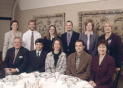
Left to right. Top: Sarah Caddick, David Hutcheson, Rebecca Sappington,
David Calkins, Martha Morehouse, Rita Loskill.
Seated: Patrick Hines, Nicholas Marsh-Armstrong, Monica Vetter,
Philip Horner, Kathleen Gwynn.
|
CFC Annual Meeting, Year Two
On December 3rd and 4th, the Kirsch Foundation and the Glaucoma Research Foundation (GRF) hosted the second CFC annual review meeting in San Francisco, California. It was a very hopeful meeting. CFC members have established a rodent model and determined that it actually develops glaucoma, not just the symptoms of the disease. As a result, in 2004, the investigators plan to use the colony to identify potential genes that predispose someone to the disease or actually cause the disease to develop. Both Kirsch Foundation and GRF staff and Board are excited by the tremendous enthusiasm the investigators demonstrate for the project, their long-term commitment to this research initiative and the progress that is being made to find a cure.
Second Year Progress Report from the CFC
The past year has been a period of significant progress. The four labs met four times over the course of 2003 to discuss progress and design new experiments. In addition, the consortium held monthly conference calls to keep abreast of new data and ideas. Through these interactions, the consortium established priorities as a group and defined key experiments that will provide new insight into how glaucoma progresses. In the last year the consortium has gone from intellectually collaborating and training to physically implementing large-scale research projects. The results and interpretation of these projects will unfold in 2004.
Progress on the establishment of the rodent model of glaucoma as well as each of the four objectives identified at the beginning of the collaborative research effort is listed below:
- Animal model of glaucoma for experimental analysis: A primary goal of the consortium was to work with an animal model of glaucoma that would permit experimental investigation into the mechanisms of disease progression. The DBA/2J mouse strain is an inbred mouse line available through Jackson Labs that has been shown to develop glaucoma in an age-dependent manner, showing many of the hallmark features seen in human glaucoma. It was necessary to establish a centralized animal core to breed and monitor these mice so that they would be available to all labs in the consortium for experimental analysis. In 2003, the Horner lab recruited an animal care technician to care for the breeding and characterization of the DBA/2J mouse model of glaucoma. The colony is now comprised of nine groups of mice that are staged at 1 through 12 months of age in order to maintain the needed groups for the proposed research. In 2003, the Horner lab experimented with three techniques to monitor the intraocular pressure (IOP) of DBA/2J mice and developed a database of IOP measurements to be used for properly staging the disease. Tissue from the colony has already been disseminated to consortium labs and this process will be accelerated in 2004. The current year plan also requires the development of a new DBA/2J mouse model, which will allow the real time visualization of the disease and provide a new tool to the greater glaucoma research community.
- Objective 1 - Assessing the repair response in glaucoma: One goal of the consortium is to assess whether retinal ganglion cell loss during glaucoma progression triggers proliferation of a retinal stem cell population leading to retinal repair. In 2003 the consortium completed an analysis of stem cell activity in the adult mouse retina. The group’s findings indicate that stem cells are not active during the progression of glaucoma and that the disease may actually repress amplification of stem cells. This would indicate that the retina is unique compared to other areas of the brain where scientists have found that stem cells respond to neurodegeneration. In the coming year the consortium will move to understand how repression of stem cell repair is mediated and explore methods to activate repair or replacement of lost neurons by stem cells.
- Objective 2 - Profiling changes in gene expression during glaucoma progression by micro array analysis: In order to understand the mechanisms underlying disease progression in glaucoma it is necessary to determine how retinal ganglion cells are changing before they actually die. This may allow the research team to understand why these cells die and ultimately enable them to intervene in the disease before retinal ganglion cells are lost. The CFC researchers therefore plan to profile changes in gene expression in retinal ganglion cells during glaucoma progression using micro array technology. This technology allows researchers to simultaneously monitor changes in the expression of thousands of genes, and compile a fairly complete profile of retinal ganglion cells at various stages of the disease. In 2003, the Vetter lab established all of the necessary tools and methods needed for this study and then tested its ability to perform the analysis on normal mouse retina. In 2004, the lab will complete an initial analysis of retinal ganglion cells in the DBA/2J mouse model of glaucoma and implicate candidate genes in glaucoma progression. CFC researchers will then design experiments to test the role of these genes in retinal ganglion cell loss.
- Objective 3 - Developing tools to target gene expression to specific cell populations in the eye: An important goal of the consortium is to target gene expression to retinal ganglion cells or retinal progenitor cells to allow the investigators to test hypotheses about the mechanisms underlying glaucoma and potentially intervene in disease progression. During this past year the consortium met one of the goals initially set, namely to identify regulatory regions that drive expression in retina progenitor cells. This was accomplished by taking advantage of reagents generated by the human genome project and the ability to screen large transgenes in the frog. The candidate gene on which the team first focused was a gene by the name of Frizzled-5, which belongs to a class of molecules that are important for multiple aspects of development. The Vetter lab had evidence that this gene was expressed in retina progenitors in both mice and frogs, making it an optimal candidate for analysis. The Marsh-Armstrong lab obtained a large piece of human DNA containing this gene and surrounding DNA, inserted a fluorescence reporter gene into the human Frizzled-5 gene by procedure called recombineering, and made transgenic frogs with this engineered DNA. The Vetter and Marsh-Armstrong labs then collaborated again in order to demonstrate that the frogs developed expression of the fluorescent reporter gene in the retina progenitor cells. In light of the exciting results from the Horner and Calkins laboratories, the goal for 2004 will be to meet another goal the consortium set out, namely to find regulatory regions that drive specific expression in retinal ganglion cells. These regulatory regions will be particularly useful in efforts to try to alter disease progression in the DBA/2J mice.
- Objective 4 - Assessing neuronal/glial interactions during glaucoma progression: The goal of this strategy is to determine the interrelationship between neuronal and glial cell loss during the early stages of glaucoma to gain insights into the mechanisms underlying disease progression. This strategy has been greatly expanded in its scope and pace of accomplishment by involving all members of the consortium. The characterization of molecular and anatomical changes in glial and neurons in the DBA/2J mouse model of glaucoma has become a central goal of the group. It is clear that a linear analysis of early changes in the DBA/2J mouse could open new avenues of research and identify novel precipitating events that induce disease progression. In 2003, all consortium labs have shared a longitudinal sample of retinas or isolated neurons and glia from the glaucoma mouse mode. The Calkins lab has quantitatively assessed neuronal loss and verified that the mouse model closely resembles the human disease. All CFC laboratories are now quantitatively assessing large amounts of tissue from diseased retinas. It is too early to draw conclusions but a complete assessment of this data set will be accomplished in the first part of 2004. The information should reveal the earliest cellular changes in the disease, that is, changes that occur before the loss of ganglion cells. This information will help to identify novel therapeutic targets.
Our progress in Year Two (2003) has shown that the four participating labs can effectively combine forces to tackle fundamental questions about glaucoma:
- We have made significant progress in establishing an animal core to provide mice that develop glaucoma for experimental analysis.
- We have begun to characterize disease progression in these mice and have detailed analysis of cellular changes in neuronal and glial cells.
- We have laid the groundwork for a comprehensive molecular analysis of this disease and have begun to develop tools for targeting specific cell populations in the retina to allow us to intervene in glaucoma progression.
The coming year (2004) shows promise of bringing important new insights into the mechanisms of glaucoma progression and identifying potential clinical targets.
Three Year Progress Report from the CFC
The Catalyst for a Cure consortium is different from most research efforts in glaucoma, both by its design and its intent. The CFC was formed in 2002 by four investigative groups chosen by our scientific advisory board for their particular expertise in neurobiology, ophthalmology and developmental genetics. Each group forms a nucleus whose purpose is to facilitate the rapid and efficient development of technologies pertinent to understanding the causes of glaucoma and identifying potential new treatments. The intent was simple: to foster a fresh understanding of glaucoma from a decidedly "non-ophthalmic" viewpoint. With an investigator in spinal cord repair (Phil Horner, University of Washington), two in developmental neurobiology (Monica Vetter, University of Utah, and Nick Marsh-Armstrong, Johns Hopkins University), and a fourth in retinal physiology and genetics (David Calkins, Vanderbilt University), the field was set for the CFC to develop a novel approach to understanding how glaucoma blinds. Increased intraocular pressure in the front of the eye – so endemic in glaucoma – produces mechanical stress on the optic nerve and retina, which results in the death of the ganglion cells and their fibers. The CFC therefore focuses on understanding the signals that cause ganglion cells to die and on developing new ways to slow the loss of optic nerve fibers.
In its first three years, the CFC has made progress on several fronts. The team has found that pressure-induced mechanical stress on the retina induces inflammatory signals that contribute to the pattern of degeneration that eventually leads to blindness. This inflammation involves interactions between the ganglion cells and other cells called "glia," which normally provide immunity from pathogens carried in the retinal blood supply. The CFC believes that hope for abating the spread of ganglion cell loss may lie not only in inhibiting the inflammatory signals between ganglion cells and glia, but in promoting and exploiting the protective signals that glia provide. Much of this protection is active during the development of the retina, so the CFC is intent on understanding the genetic cues that control the growth of ganglion cells during this critical period. Already members of the CFC have identified certain factors that control how ganglion cells develop and are working to exploit these factors to impede glaucoma.
The team has also focused on applying the latest technologies in "high-throughput" genetic testing. The retina, like the rest of the brain, is composed of many different kinds of cells and connections, and traditional means of genetic testing generally do not allow the sort of resolution necessary to isolate only those changes occurring in ganglion cells. The CFC worked diligently early on to develop a technique to isolate ganglion cells from the rest of the retina and to extract the genetic material from those isolated cells. The CFC then was able to use printed microchips on which thousands of genes are represented to compare changes in genetic patterns during disease progression. The resulting information has been useful for pinpointing the biochemical cascades involved in ganglion cell death. Indeed one of the most prominent results so far indicates that retinal ganglion cells are unique in the brain because they express particular genes that are susceptible to changes in pressure. Another important finding is that ganglion cells in glaucoma resemble the neurons in the brain that die in other diseases, like Alzheimer's and Parkinson's disease. This means that the knowledge gained by studying glaucoma might be helpful in understanding other diseases of the brain and vice versa.
The CFC believes that by bringing to bear new tools that are not generally associated with glaucoma research they will be able to shed light on potentially new avenues of therapeutic intervention. The results from the first three years indicate that so-called "death signals" in glaucoma occur very early in the disease, perhaps even earlier than loss in vision can be detected. Thus, in the next three-year phase, the team will work on better understanding the mechanisms of these earlier pathological changes and on genetic means to alter these mechanisms. With this information, the team will then work to identify proteins and genes that have the greatest potential as targets for new drugs. Finally, the CFC hopes to apply this information to intervene during the early stages of the disease in one of its multiple experimental models. Thus, at the completion of the second phase, the CFC hopes to have shed considerable light on the possibilities of new therapeutic systems for glaucoma. Like the CFC focus, the anticipation is that new therapies will center upon neurological factors and therefore may have applicability to other diseases of the central nervous system.

Left to right: David Calkins, Philip Horner, Nick Marsh-Armstrong, Monica Vetter
|
|
|